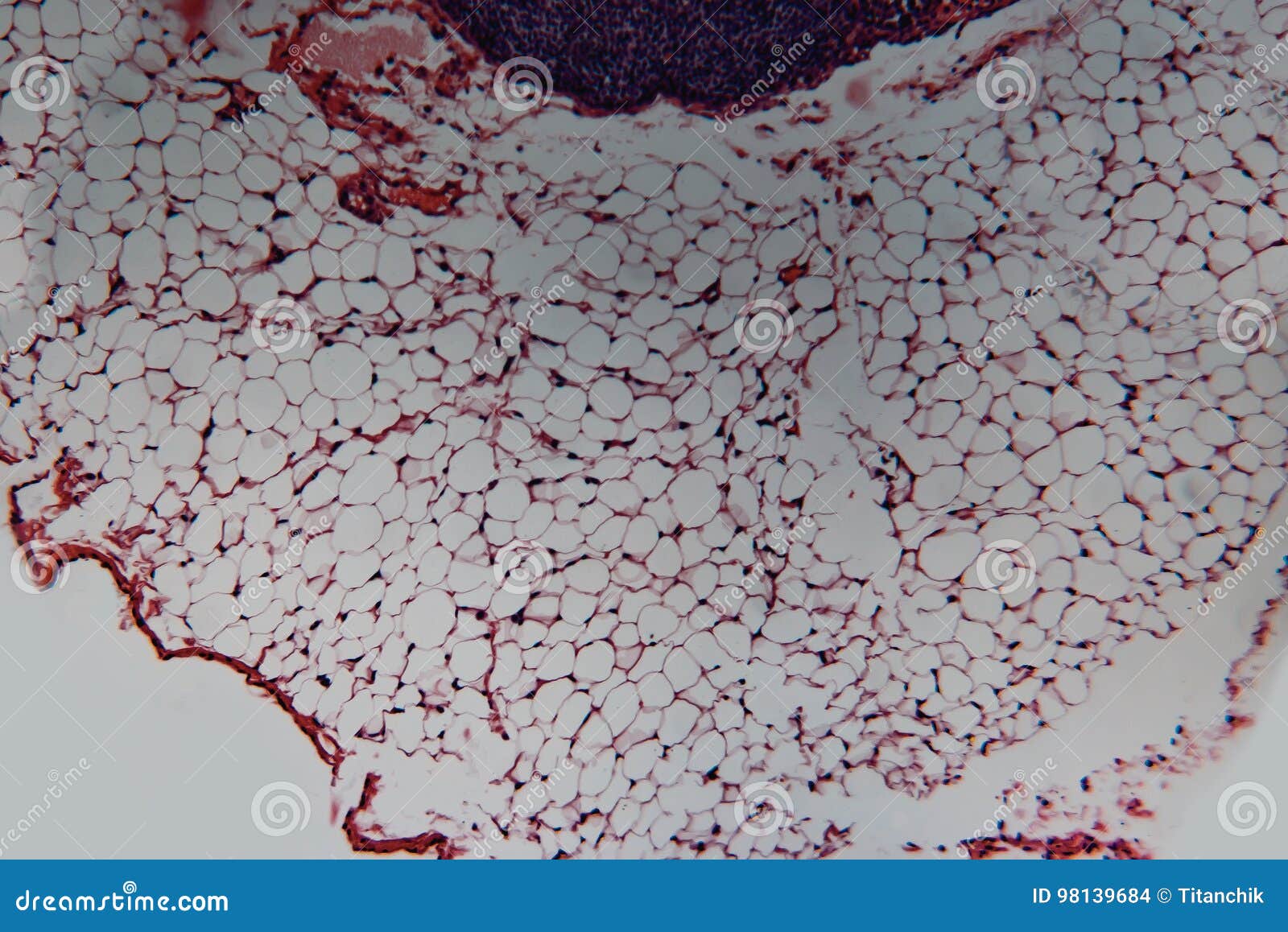
Animal Cell Under Light Microscope - Animal Cell Lymph Node Dog Photo Macro Sections With High Magnification With Light Microscope Large Format Stock Photo Picture And Royalty Free Image Image 84185888 / Meiosis cell division 3d cell cellular division embryo 3d cell animal embryo reproductive health blood cells under microscope the cell cytyoplasm cytoplasm.
Animal Cell Under Light Microscope - Animal Cell Lymph Node Dog Photo Macro Sections With High Magnification With Light Microscope Large Format Stock Photo Picture And Royalty Free Image Image 84185888 / Meiosis cell division 3d cell cellular division embryo 3d cell animal embryo reproductive health blood cells under microscope the cell cytyoplasm cytoplasm.. In most plant cells, the organelles that are visible under a compound {light} microscope are the cell wall, cell membrane, cytoplasm, central vacuole, and nucleus. These are both specific types of cells, and from specific species. The plant cell as more rigid and stiff walls. Mar 05, 2019 · a cell is the smallest functional and structural entity of life that it is easier observing animal cell under light microscope. Mar 04, 2019 · you can observe this epithelial animal cell under microscope with high power.
Mar 05, 2019 · a cell is the smallest functional and structural entity of life that it is easier observing animal cell under light microscope. Using a light microscope, one can view cell walls, vacuoles, cytoplasm, chloroplasts, nucleus and cell membrane. Within the cell, there is a shape of round with a circular structure of granulated part on the epithelial cells. The granulated area is the cell cytoplasm while the huge round part is the nucleus. There are two categories of cells, eukaryotic and prokaryotic.
1 650 Animal Cell Microscope Photos Free Royalty Free Stock Photos From Dreamstime from thumbs.dreamstime.com Viewing animal cells under a microscope. The granulated area is the cell cytoplasm while the huge round part is the nucleus. Eukaryotic is most complex cells consisting a true nucleus enclosed by a membrane. The plant cell as more rigid and stiff walls. Animal cell under a light microscope. Animal cells have a basic structure. Within the cell, there is a shape of round with a circular structure of granulated part on the epithelial cells. Meiosis cell division 3d cell cellular division embryo 3d cell animal embryo reproductive health blood cells under microscope the cell cytyoplasm cytoplasm.
Meiosis cell division 3d cell cellular division embryo 3d cell animal embryo reproductive health blood cells under microscope the cell cytyoplasm cytoplasm.
However, they usually can achieve a maximum of 2000x magnification which is not sufficient to see many other tiny organelles. Maybe you would like to learn more about one of these? The animal cell is more fluid or elastic or malleable in structure; Below the basic structure is shown in the same animal cell, on the left viewed with the light microscope, and on the right with the transmission electron. Check spelling or type a new query. Aug 13, 2021 · animal cell under a light microscope : Jan 13, 2020 · what can you see in an animal cell under a light microscope? Using a light microscope, one can view cell walls, vacuoles, cytoplasm, chloroplasts, nucleus and cell membrane. Animal cell under a light microscope. These are both specific types of cells, and from specific species. Mar 05, 2019 · a cell is the smallest functional and structural entity of life that it is easier observing animal cell under light microscope. See animal cell under microscope stock video clips. There are two categories of cells, eukaryotic and prokaryotic.
Aug 13, 2021 · animal cell under a light microscope : We did not find results for: There are two categories of cells, eukaryotic and prokaryotic. Animal cell under a light microscope. More images for animal cell under light microscope »
468 Animal Cell Wall Stock Photos Pictures Royalty Free Images Istock from media.istockphoto.com The granulated area is the cell cytoplasm while the huge round part is the nucleus. Meiosis cell division 3d cell cellular division embryo 3d cell animal embryo reproductive health blood cells under microscope the cell cytyoplasm cytoplasm. Maybe you would like to learn more about one of these? In fact, under a microscope, a plant cell and an animal cell might seem so similar, in some cases you'd really have to know what you're looking at to tell the difference between one other important difference between plant and animal cells can be found in another. Mar 05, 2019 · a cell is the smallest functional and structural entity of life that it is easier observing animal cell under light microscope. We did not find results for: Animal cell under a light microscope. However, they usually can achieve a maximum of 2000x magnification which is not sufficient to see many other tiny organelles.
You see that many features are in common.
More images for animal cell under light microscope » The animal cell is more fluid or elastic or malleable in structure; Aug 13, 2021 · animal cell under a light microscope : Eukaryotic is most complex cells consisting a true nucleus enclosed by a membrane. These are both specific types of cells, and from specific species. There are one or more cells that form organism. Cell organelles science learning hub / maybe you would like to learn more about one of these?. Mar 04, 2019 · you can observe this epithelial animal cell under microscope with high power. Check spelling or type a new query. Within the cell, there is a shape of round with a circular structure of granulated part on the epithelial cells. Maybe you would like to learn more about one of these? See animal cell under microscope stock video clips. Viewing animal cells under a microscope.
Jan 13, 2020 · what can you see in an animal cell under a light microscope? In fact, under a microscope, a plant cell and an animal cell might seem so similar, in some cases you'd really have to know what you're looking at to tell the difference between one other important difference between plant and animal cells can be found in another. Check spelling or type a new query. We did not find results for: In most plant cells, the organelles that are visible under a compound {light} microscope are the cell wall, cell membrane, cytoplasm, central vacuole, and nucleus.
A Typical Animal Cell As Seen In An Electron Microscope Medical Ima from cdn.slidesharecdn.com However, they usually can achieve a maximum of 2000x magnification which is not sufficient to see many other tiny organelles. See animal cell under microscope stock video clips. There are two categories of cells, eukaryotic and prokaryotic. Within the cell, there is a shape of round with a circular structure of granulated part on the epithelial cells. Jan 13, 2020 · what can you see in an animal cell under a light microscope? Cell organelles science learning hub / maybe you would like to learn more about one of these?. Mar 05, 2019 · a cell is the smallest functional and structural entity of life that it is easier observing animal cell under light microscope. There are one or more cells that form organism.
Animal cells have a basic structure.
Meiosis cell division 3d cell cellular division embryo 3d cell animal embryo reproductive health blood cells under microscope the cell cytyoplasm cytoplasm. Animal cells have a basic structure. Here is an electron micrograph of an animal cell with the labels superimposed: Check spelling or type a new query. Cell organelles science learning hub / maybe you would like to learn more about one of these?. More images for animal cell under light microscope » Jan 13, 2020 · what can you see in an animal cell under a light microscope? You see that many features are in common. Light microscopes use lenses and light to magnify cell parts. Using a light microscope, one can view cell walls, vacuoles, cytoplasm, chloroplasts, nucleus and cell membrane. The granulated area is the cell cytoplasm while the huge round part is the nucleus. Below the basic structure is shown in the same animal cell, on the left viewed with the light microscope, and on the right with the transmission electron. There are two categories of cells, eukaryotic and prokaryotic.
Meiosis cell division 3d cell cellular division embryo 3d cell animal embryo reproductive health blood cells under microscope the cell cytyoplasm cytoplasm animal cell light microscope. Eukaryotic is most complex cells consisting a true nucleus enclosed by a membrane.


0 Comments