Animal Cell Labeled Microtubules / A Labeled Diagram of the Animal Cell and its Organelles - Tubulin labeling in live cells with minimal cytotoxicity.. Conduit pt et al., centrosome function and assembly in animal cells. Both of these structures are found in animal cells, but not plant cells. During cell division (mitosis), the centrosome divides and the two parts move to opposite sides of the dividing cell. One of the primary concerns with using fluorescent taxanes to label microtubules is the possibility of cytotoxicity upon continuous incubation. Microtubules as their name implies, microtubules are small hollow tubes.
Microtubules are exactly how they sound: Parts of an animal cell biography. Cell biologist thomas maresca and senior research fellow vikash verma at the university of massachusetts amherst say they have, for the first time, directly observed and recorded in animal cells a pathway called branching microtubule nucleation, a mechanism in cell division that had been. They are microscopic hollow tubes found inside cells that also provide motor functions for the. Picone r, ren x, ivanovitch kd, clarke jdw, mckendry ra, baum b (2010) a polarised population of dynamic microtubules mediates homeostatic length control in animal cells.
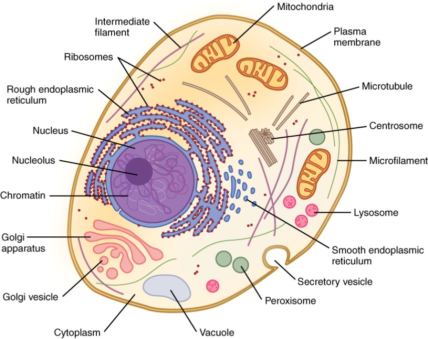
Animal Cell Parts and Functions - WithCarbon from image.jimcdn.com In most mammalian cells, microtubules form a tube of 13 protofilaments. Microfilaments bear a resemblance to microtubules but microfilaments are softer and smaller in diameter. These straight, hollow cylinders are found throughout the cytoplasm of all. Animal cells are of various sizes and have irregular shapes. An animal cell ranges in size from 10 to 30 µm. Cilia are projections from a cell that can m. They help the cell resist compression, provide a track. In animal cells, microtubules radiate outwards from an organelle in the center of the cell called a centrosome, which is a microtubule organizing center (mtoc).
Microtubules in the cell consist of microscopic structures formed in hollow tubes and constructed in a series of linear rings.
Animal cells are unique in that they contain special organelles for the construction and maintenance of microtubules, organelles known as centrioles. Animal cells from the basic structural units of all tissues and organs of the body. Microfilaments are organelle cells formed from actin and myosin proteins. In animal cells, microtubules radiate outwards from an organelle in the center of the cell called a centrosome, which is a microtubule organizing center (mtoc). Microbodies microtubules and microfilament are the additional cell organelles. Two or more centrioles will aggregate perpendicularly to each other. Microtubules also form cell structures called centrioles and asters. The walls of the microtubule are made of with a diameter of about 25 nm, microtubules are the widest components of the cytoskeleton. Although centrosomes with centrioles may assist the organization of microtubule construction in animal cells, they are not crucial for this particular function in all eukaryotes; This procedure compromises neither the polymerizability of the proteins nor their affinities for unlabeled proteins. Animal cells are of various sizes and have irregular shapes. Under the microscope, an animal cell shows many different parts called organelles, that work together to keep. Essentially, the two aggregated centrioles act as anchors for the rest of the cell's.
The fungi and the majority of plant cells lack centrosomes. Animal cells are unique in that they contain special organelles for the construction and maintenance of microtubules, organelles known as centrioles. The centrosome is where microtubules are made. Under the microscope, an animal cell shows many different parts called organelles, that work together to keep. In most mammalian cells, microtubules form a tube of 13 protofilaments.
Unlabeled Animal Cell - ClipArt Best - ClipArt Best from www.clipartbest.com Two or more centrioles will aggregate perpendicularly to each other. Microscopic hollow tubes found inside eukaryotic cells and some prokaryotic bacteria cells that provide structure and motor functions. Microtubule proteins and tubulin have been purified from brain and labeled with dichlorotriazinyl fluorescein (dtaf). Microtubules as their name implies, microtubules are small hollow tubes. In most mammalian cells, microtubules form a tube of 13 protofilaments. Also cilia and flagella are made of microtubules. This procedure compromises neither the polymerizability of the proteins nor their affinities for unlabeled proteins. During cell division (mitosis), the centrosome divides and the two parts move to opposite sides of the dividing cell.
Microscopic hollow tubes found inside eukaryotic cells and some prokaryotic bacteria cells that provide structure and motor functions.
Microtubules are exactly how they sound: The conventional mtoc in animal cells is the centrosome, an. Microfilaments bear a resemblance to microtubules but microfilaments are softer and smaller in diameter. Two or more centrioles will aggregate perpendicularly to each other. Animal cells are common names for eukaryotic cells that make up animal tissue. Also cilia and flagella are made of microtubules. These hollow rods help give structure and shape to the cell. Microtubules are polymers of tubulin that form part of the cytoskeleton and provide structure and shape to eukaryotic cells. One of the primary concerns with using fluorescent taxanes to label microtubules is the possibility of cytotoxicity upon continuous incubation. This note contains detail information about microbodies microtubules peroxisomes are the microbodies found in many animal cells and in a wide range of plants. Microscopic hollow tubes found inside eukaryotic cells and some prokaryotic bacteria cells that provide structure and motor functions. Microtubules also form cell structures called centrioles and asters. Conduit pt et al., centrosome function and assembly in animal cells.
Microtubule proteins and tubulin have been purified from brain and labeled with dichlorotriazinyl fluorescein (dtaf). Our body starts its existence at fertilization from a single cell, the diploid zygote. In animal cells, microtubules radiate outwards from an organelle in the center of the cell called a centrosome, which is a microtubule organizing center (mtoc). The nucleus of a cell contains its dna and controls the cell's growth and. Microtubules are found in the cytoplasm of all types of eukaryotic cells with rare absence, such as in human erythrocytes.
Animal Cell Model Diagram Project Parts Structure Labeled ... from lh3.googleusercontent.com In animal cells, microtubules radiate outwards from an organelle in the center of the cell called a centrosome, which is a microtubule organizing center (mtoc). They help the cell resist compression, provide a track. Microtubules as their name implies, microtubules are small hollow tubes. Microtubules are polymers of tubulin that form part of the cytoskeleton and provide structure and shape to eukaryotic cells. Animal cells are unique in that they contain special organelles for the construction and maintenance of microtubules, organelles known as centrioles. Our body starts its existence at fertilization from a single cell, the diploid zygote. Most of the cells size range between 1 illustrated in figure 2 is a pair of fibroblast deer skin cells that have been labeled with fluorescent probes microtubules. The cellular organization of microtubules varies between cell types, but in most cells, the minus ends of microtubules are anchored to the centrosomes near the nucleus while the plus ends radiate towards the periphery of the cell.
Mtocs allow the cell to control where and when to assemble microtubules.
A simple, quantitative model of cellular extension driven by microtubules recapitulates cell elongation on lines, the citation: This procedure compromises neither the polymerizability of the proteins nor their affinities for unlabeled proteins. The conventional mtoc in animal cells is the centrosome, an. Microtubules involved in movement of the cell (cilia and flagella and the basal bodies that connect them to the cell) very stable and not dynamic like the. Also cilia and flagella are made of microtubules. Microtubules are polymers of tubulin that form part of the cytoskeleton and provide structure and shape to eukaryotic cells. Nat rev mol cell biol 2015; These hollow rods help give structure and shape to the cell. The nucleus of a cell contains its dna and controls the cell's growth and. Organization and shape of animal cells, intracellular organelle mobility, maintain axons, spindle formation. Animal cells are common names for eukaryotic cells that make up animal tissue. Animal cells are of various sizes and have irregular shapes. Picone r, ren x, ivanovitch kd, clarke jdw, mckendry ra, baum b (2010) a polarised population of dynamic microtubules mediates homeostatic length control in animal cells.


0 Comments